How The Thyroid and Parathyroid Surgery Is performed?
WHAT IS GOITER?
The enlargement of the thyroid gland is called goitre. The thyroid is located in the anterior-inferior part of the neck, on both sides of the trachea. It synthesises thyroid hormone, which plays a vital role in regulating metabolism.
Goitre can be:
- Simple or nodular, depending on whether the gland is enlarged, it is associated or not with the presence of nodules, cystic or solid.
- Normally functioning, hypo functioning or hyperfunctioning (toxic), depending on whether the thyroid function is normal, decreased or increased.
SYMPTOMS OF GOITER
Most patients are asymptomatic at the time of diagnosis, usually discovered incidentally during a physical examination performed for another reason.
On other occasions, the patient goes to his doctor to notice the appearance of a lump or tumour of variable size on the anterior face of the neck, which may or may not be painful on palpation.
There are other rarer symptoms related to compression of the gland on neighbouring structures.
DIAGNOSIS OF GOITER
- Anamnesis: the patient should be asked about the evolution time and changes in the size of the goitre (in general, long-standing goitres suggest benignity, while rapidly growing ones suggest malignancy). Exposure to cervical radiation (increases the risk of thyroid cancer), a history of autoimmune disease and the presence of compressive symptoms.
- Physical examination: Palpation to determine the size, shape, and consistency of the thyroid. The normal thyroid gland is not visible, and on palpation, it is elastic in texture and small in size.
- Blood tests: the blood measurement of the hormones involved in thyroid function (TSH, T4) helps us to know if there is normal, increased or decreased function. Most of the time, thyroid function is normal. You can also see in the blood test the existence or not of antibodies against the thyroid (their presence points to an immunological cause) or other substances such as calcitonin, which points to a cancerous origin of the goitre.
- Simple x-ray: it helps us to see if there is tracheal displacement, calcifications in the gland or undiagnosed lung lesions.
- Thyroid ultrasound: it is the technique of choice for the study of thyroid morphology since it allows for defining the existence of nodules, their size, and whether they are solid or cystic; however, it does not provide information on the functional activity of the nodules; therefore, it does not report their benign or malignant nature. It also allows controlling the size of known nodules over time to see their evolution or guide other techniques such as thyroid puncture.
- Fine-needle aspiration puncture (FNA): allows, without the need for surgery, to determine the benign or malignant nature of a nodule. The PAAF allows obtaining thyroid cells later studied in the laboratory, thus seeing if they are benign or malignant. It is a safe technique and has few complications, being the basis for diagnosing thyroid nodules.
- Thyroid scintigraphy: this allows us to carry out a functional study of the thyroid. Thus, thyroid nodules may be hyper- uptake or hot, normal- uptake or warm, or hypo-uptake or cold. Currently, scintigraphy has been replaced by FNA (Fine-needle aspiration puncture) to determine the nature of the lesions.
TREATMENT OF GOITER
Treatment will depend on the patient’s symptoms, the size of the nodules, thyroid function, and the result of the FNA.
- If the goitre produces symptoms due to hyperthyroidism or compression (hoarseness, difficulty breathing or swallowing), the treatment must be surgical if there is no anaesthetic contraindication. Surgery consists of removing part of the thyroid (hemithyroidectomy) or all of it (total thyroidectomy).
- In very elderly or high surgical risk symptomatic patients, the treatment of choice is using radioactive iodine. This treatment usually reduces the size of the goitre by 40%.
- When the goitre is asymptomatic, the treatment will depend on the characteristics of the nodules. If the nodules are benign in appearance and smaller than 1 cm, follow-up can be done. If they are larger than 1 cm, it is advisable to biopsy them to confirm benignity. When the biopsy is suspicious of malignancy, the recommended treatment is surgery.
Follow-up consists of performing a physical examination, laboratory tests, and ultrasound to confirm that there are no relevant changes. Follow-up can be done every 6-12 months, depending on each patient.
WHAT DOES THYROIDECTOMY CONSIST OF, AND WHAT ARE ITS RISKS?
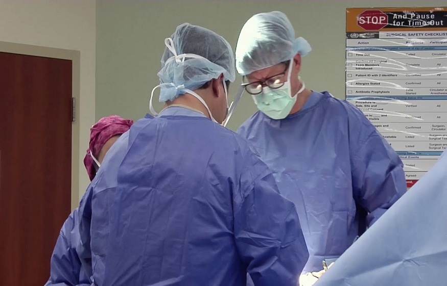
Thyroid surgery removes the gland partially (hemithyroidectomy) or completely (total thyroidectomy). The standardised access route consists of a 5-7 cm incision in the lower area of the neck, over the gland. Other less invasive access routes have been described, but few surgeons apply them today.
With the new technology, surgical times and complications have been significantly reduced. Each hemithyroidectomy usually takes 35-40 minutes. The patient is usually discharged within 24-48 hours. When the entire gland is removed, blood calcium values should be measured postoperatively. Some surgeons leave drains that are typically removed after 24 hours.
Complications are rare and are usually related to the anatomical structures surrounding the thyroid:
- Bleeding from a small blood vessel.
- Temporal dysphonia due to recurrent laryngeal nerve involvement. Exceptionally rarely, there may be permanent aphonia.
- Hypocalcaemia due to temporary involvement of the parathyroid glands. These four glands are located next to the thyroid, each the size of a lentil. Although it is enough to have one of them to fulfil its metabolic function, they must be respected to reduce the risk of hypocalcaemia.
WHAT IS PRIMARY HYPERPARATHYROIDISM?
“The excess of this hormone is related to the loss of bone tissue, which brings an increased risk of bone fracture and kidney damage, associated with the formation of kidney stones and even calcium deposits in the kidney parenchyma.”
At the neuropsychiatric level, this pathology produces a feeling of fatigue, tiredness and irritation, in addition to causing depression, dementia or aggravation of a previous state of dementia.
DIAGNOSIS OF PRIMARY HYPERPARATHYROIDISM
In most cases, the disease is diagnosed in the preclinical phase, that is, before symptoms appear, through a routine blood test performed for any other reason, in which an increase in calcium levels is detected. Once the diagnostic suspicion of hyperparathyroidism is confirmed, “starting from the premise that 90% of patients have only one diseased gland, we carry out tests to locate the adenoma, such as sestamibi scintigraphy or an ultrasound “, pointed out this expert in endocrine surgery.
TREATMENT OF PRIMARY HYPERPARATHYROIDISM
Surgery is the TREATMENT of choice in patients with primary hyperparathyroidism, with a cure rate of between 95 and 98% of cases in the first intervention performed by an expert surgeon. “The use of minimally invasive techniques increasingly focused on a selective approach, as well as the more excellent training of professionals, makes this surgery a safe and effective treatment with less postoperative pain and earlier incorporation into your daily life.